![]()
![]()
![]()
|
|
|
CRUISE REPORT
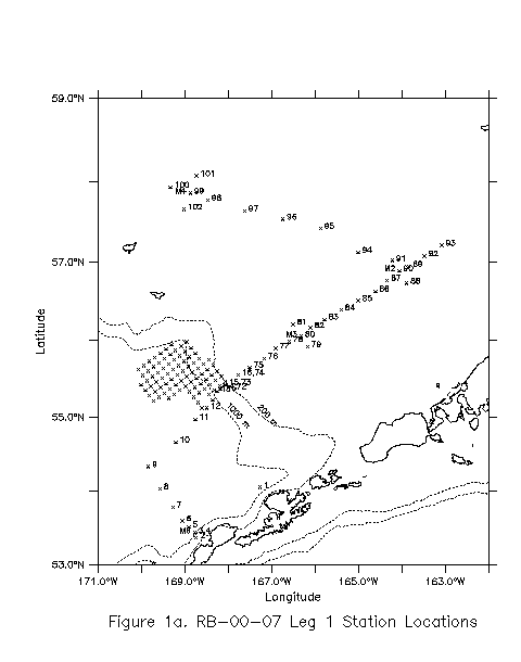
Section 2.0 - Cruise Narrative Section 3.0 - Physical and Chemical Oceanographic Measurements Section 4.0 - Bio-Optical Characterization of Bering Sea Waters Section 5.0 - Productivity Studies Section 6.0 - Acknowledgements Ship:NOAA Ship RONALD H. BROWNArea of Operations:Eastern Bering SeaItinerary:27 Aug 2000 depart Dutch Harbor, AK14 Sep 2000 arrive Dutch Harbor, AK Chief Scientist:Edward D. Cokelet, Ph DNOAA/PMEL 7600 Sand Point Way NE Seattle, WA 98115-6439 Phone: (206) 526-6820 Fax: (206) 526-6485 E-mail: Edward.D.Cokelet@noaa.gov 1.0 Background1.1 Program DescriptionThis cruise was part of the Fisheries Oceanography Coordinated Investigations (FOCI) - an effort by NOAA and academic scientists to understand the physical and biological processes that determine recruitment variability of commercially valuable fin-fish and shellfish stocks in Alaskan waters. FOCI consists of several projects including the present ones funded by the Southeast Bering Sea Carrying Capacity (SEBSCC) and the North Pacific Marine Research (NPMR) Programs.1.2 Cruise ObjectivesThe SEBSCC objective is to monitor the water properties and circulation along an oft-repeated oceanographic section in the Bering Sea starting at Mooring 6 and proceeding across the basin, onto the continental shelf past Moorings 3 and 2, and along the 70-m isobath to Mooring 4 (Fig. 1a).The purpose of the NPMR project is to understand the influence of mesoscale eddies on continental slope-shelf exchange in the Southeastern Bering Sea. The objectives are to (1) Detect movements of nutrient-rich slope water onto the shelf and relate them to temporal and spatial variations in chlorophyll, (2) Identify physical mechanisms that create slope-water fluxes onto the continental shelf, (3) Detect ocean-color variability in relation to physical processes, (4) Use shipboard measurements of near-surface optical and biological parameters to validate and extend bio-optical algorithms for use in autonomous sampling and remote sensing, and (5) Investigate the effects of on-shelf flow on phytoplankton biology. 1.3 Operating AreaSoutheastern Bering Sea (Fig. 1a).1.4 Participating OrganizationsNOAA/Pacific Marine Environmental Laboratory (PMEL)7600 Sand Point Way NE Seattle, WA 98115-6439 University of Alaska-Fairbanks (UAF) Dalhousie University (Dal) Smithsonian Environmental Research Center (SERC) 1.5 Personnel
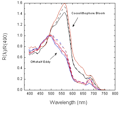
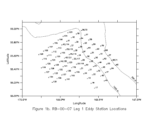
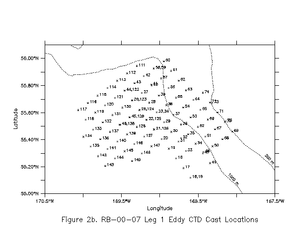
CTD Watches: Salo and Kinoshita midnight-noon (ADT = GMT - 8 hr) 2.0 Cruise NarrativeThe ship departed Dutch Harbor on 27 August after delaying one day to await the arrival of an electrical cable for the Dalhousie floc-camera system. (Owing to customs delays, the cable did not arrive on time, and we eventually left port without it.) We deployed a satellite-tracked drifting buoy in 1000 m of water north of Dutch Harbor (stn. 1, Fig. 1a and Table 1 - the Master Scientific Log). Then we proceeded to the SW end of the SEBSCC line and began CTD operations near Mooring 6 (stn. 2, Fig. 1a). CTD operations continued to station 16 on the continental shelf. (Note that because more than one CTD cast can be taken at an oceanographic station, the station numbering of Fig. 1 differs from the cast numbering of Fig. 2.)After station 16 the ship left the SEBSCC line and proceeded to the slope-shelf-exchange study area. Two separate tools provided a way to locate eddies there - sea-surface altimetry observations from the TOPEX/Poseidon satellite and satellite-tracked drifting buoy trajectories. The former were available from World Wide Web pages http://www-ccar.colorado.edu/~leben/research.html provided by Dr. Robert Leben at the University of Colorado which we were able to download throughout the cruise via Inmarsat-A satellite-telephone modem connections. Unfortunately the altimetry showed only weak eddy activity and suggested no likely site for our study. On the other hand, the trajectory of a satellite-tracked drifter drogued at 40 m and launched a few months earlier showed a cyclonic eddy of ~50 km diameter off the continental shelf. We opted to study this eddy for two reasons: (1) it was potentially near enough to the continental shelf to interact with it and push basin water landward, and (2) it rotated cyclonically (counterclockwise) which is of the opposite sense to the anticyclonic eddies usually detected in the SE Bering Sea. This gave the first opportunity to study a persistent, cyclonic eddy in this region. The ship began a rectangular grid pattern of CTD casts (stations 17-71 of Fig. 1b, casts 15-75 of Fig. 2b). Temperature and salinity measurements allowed us to compute water density, dynamic height and geostrophic velocities (usually referenced to 1500 dbar) normal to CTD sections. Graphs of the transects, available within ~1 hour of transect completion, showed cyclonic eddy flow with maximum velocities of ~25 cm/s, a velocity-maximum core at ~300 m depth, and a diameter of ~80 km. We seeded the eddy with one satellite-tracked drifter that measured ocean color (a color drifter, stn. 29). After the initial slices through the eddy, we filled in the sampling grid toward the continental shelf break, looking for signs of on-shelf flow. Geostrophic velocity sections became less useful in shallow water where it was too shallow to refer them to 1500 dbar, and a level of no motion assumed to be at the sea bottom seemed untenable. After several days of sampling, we deployed two more drifters at the north (stn. 70) and south (stn. 71) ends of the shoreward side of the eddy study region. We planned to return to the drifter showing the greatest on-shelf flow and continue sampling. SEBSCC Line sampling resumed at stations 72-74 which repeated stations 14-16 of several days earlier (Fig. 1a). Mooring 3's Argos transmitter had ceased transmitting a few weeks before the cruise, and we could not locate the mooring on the surface. Attempts to enable and range upon its underwater acoustic release failed, and we presumed the mooring lost. Intensive bio-optical sampling was conducted near SEBSCC Mooring 3's deployment site (Table 1 and Fig. 1a). At dawn on 7 September near station 89, the sea was aquamarine in color indicating the presence of a coccolithophorid bloom. Owing to darkness, it was not known where that condition was first encountered, but it probably occurred somewhere between stations 83 and 89. Extra bio-optical sampling efforts were laid on to take advantage of the situation. Mooring 2 was in good condition based upon a visual inspection. We made periodic attempts to contact Mooring 3's acoustic release. Surprisingly, we got a positive response at a range of ~6 km near station 97. A search pattern gave more responses at ~2 km range, and the ship was able to range within ~200 m. The position was noted and relayed to PMEL for future mooring recovery. SEBSCC Line sampling proceeded to Mooring 4 where it was completed at stn. 102 (Fig. 1a). Then the ship returned to the eddy study area. The two drifters deployed over 3 days earlier at stns. 70 and 71 did not show much on-shelf flow, so we began sampling the seaward side of the cyclonic eddy at stn. 103 (Fig. 1b). CTD casts were taken to 1500 m filling in the 10 x 10 km grid begun earlier. Bio-optical work focussed on obtaining samples within and outside of the eddy to draw contrasts. Two drifters were deployed near the southwestern edge of the eddy at stns. 134 and 139. This work continued to the last station (stn. 141) when we returned to Dutch Harbor as scheduled. 3.0 Physical and Chemical Oceanographic MeasurementsConductivity-Temperature-Depth (CTD) casts were made with a Sea-Bird 911plus instrument with dual temperature and conductivity sensors. On the continental shelf, the instrument package carried a WETLabs WETStar fluorometer. The profiler carried a Benthos altimeter with 100 m range, and casts were to the shallower of 1500 m or 10 m above the bottom. Generally, two salinity samples were taken on each cast - one shallow and one deep - and analyzed with the ship's AutoSal for conductivity calibration purposes. CTD data were processed to 1-dbar averages.A 150-kHz RDI acoustic Doppler current profiler (ADCP) ran continuously measuring the velocity of the water relative to the ship. Auxiliary input from a Trimble P-code GPS receiver, Sperry ring-laser-gyro and Seatex Seapath 200 GPS-based attitude determination unit allow for the determination of absolute current velocity relative to the sea bottom. The ship carried a thermosalinograph to measure the temperature and salinity of near-surface waters and a flow-through Turner fluorometer to measure near-surface fluorescence on a continuous basis. Nutrient samples were collected from the majority of the CTD casts and were analyzed on board using an Alpkem RFA300 Auto-Analyzer. A total of 1032 samples were analyzed. There were 355 analyzed from the SEBSCC Line, 631 from the eddy transects and 46 for C/N productivity studies. 4.0 Bio-Optical Characterization of Bering Sea watersby Richard Davis, Margaret Davey, Catherine Brown, Yannick Huot, Kristian Curran and Jason FoxWe collected measurements of biological and optical properties of near-surface waters throughout the cruise. This section outlines some of the preliminary results and explains types of data collected. Emphasis is on aspects directly related to NPMR research. 4.1 Solar RadiationWe measured solar radiation almost continuously using a Satlantic multichannel visible detection system (MVDS). Spectral irradiance (mW cm-2 nm-1) was measured in seven wavebands between 325 and 700 nm, including the wavebands corresponding to the SeaWiFS sensor. Data were logged every 10 s. The instrument cable exhibited problems for the first days of the cruise and was subsequently repaired on board. Data are available at full resolution.4.2 Upwelling RadianceA Satlantic Tethered Spectral Radiometer Buoy (TSRB) was deployed at ~44 stations to measure upwelling radiance (Lu; mW cm-2 nm-1 sr-1) and downwelling solar irradiance (Ed; mW cm-2 nm-1) in 123 wavelength bands between 400 and 800 nm, with roughly 3.3 nm resolution. Typical TSRB deployments lasted 10-30 minutes but could exceed one hour in duration.The ratio of Lu to Ed gives the reflectance spectrum (R; sr-1) which shows the biologically determined optical variability associated with the coccolithophore bloom as encountered on the middle shelf. Figure 3 contrasts deployments inside and outside of the bloom. The reflectance spectra have been normalized to R(490).
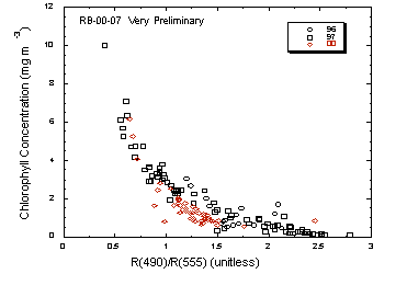
4.3 Chlorophyll aWe measured the concentration of chlorophyll a fluorometrically in triplicate for well over 200 samples. Samples were also frozen for parallel determination in the lab using fluorometric methods and HPLC. Generally, we sampled with a bucket during optical buoy measurements (depth = 0) and also from 4 depths sampled with the CTD. The frequency distribution (subject to recalibration) of surface chlorophyll concentration shows that we sampled oligotrophic and mesotrophic waters with good coverage, and also some bloom waters. We froze samples to measure the spectral absorption coefficients of particulate matter in surface waters. We also filtered water through a 0.2 mm filter and then stored it for transport to Dalhousie where we can measure the absorption due to dissolved materials.An objective of our study was to relate measures of near-surface reflectance to surface chlorophyll. These data are consistent with many studies of remote sensing that identify log(Lu(490)/Lu(555)) as a measure of chlorophyll. We report reflectance here because variability in solar Ed(490)/Ed(555) was measured, and will be incorporated into our model.
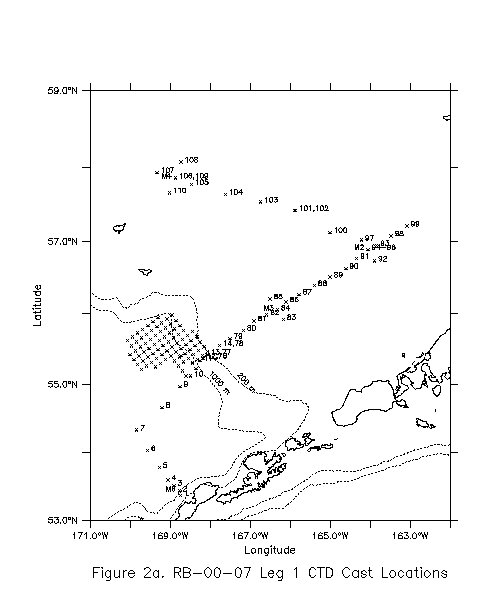
A major task of this cruise was to develop an algorithm for interpretation of optical drifter data. This requires comparison of surface chlorophyll with ocean color measurements. The preliminary analysis of Figure 4 indicates that a robust algorithm can be developed for chlorophyll concentrations > 1 mg m-3. At this time, the relationship for bluer waters looks weak. Caution is needed, however, because these data are for bucket samples only, and vertical gradients near the surface can affect the signal in clearer waters. 4.4 Penetration of Spectral IrradianceDuring almost every deployment of the tethered radiometer, we deployed a free-falling multichannel profiling radiometer, with sensors to measure downwelling irradiance in 13 wavebands (305, 323, 338, 380, 412, 443, 490, 510, 532, 555, 670, 683, and 700 nm), including those of the SeaWiFS sensors. High-quality data were obtained, which will allow us to calculate diffuse attenuation coefficients for each waveband as functions of depth for each profile. Proper analysis requires careful treatment of dark blanks, which will be done at Dalhousie. We will report diffuse attenuation coefficients at 490 nm and relate these coefficients quantitatively to upwelling radiance ratios.4.5 Bio-Optical Profiling PackageAt approximately 35 stations a bio-optical profiling package was deployed. This package contained a WETLabs ac-9, a Chelsea FASTtracker fluorometer, a HOBI Labs HydroScat-6, and a digital floc camera. Each instrument is described below. A typical deployment would include a drop to 40 m at 15 m/min descent and ascent rates. The package was recovered, a 0.2 mm filter placed in front of the inlet of the ac-9, and the package redeployed with a 15 m/min descent rate and a 5 m/min ascent rate.4.5.1 Absorption and Attenuation Absorption (a) and attenuation (c) of the upper water column were measured at 9 wavelengths (412, 440, 488, 510, 532, 555, 650, 676, 715 nm) using a WETLabs ac-9. This instrument has a 25 cm path length. Two profiles were performed at each station. The first was an unfiltered drop to measure a and c for all constituents in the water. A second filtered cast (0.2 mm) was performed so that a and c of dissolved material only was measured. Daily calibrations of the instrument were performed with ship water run through an organic-removal filter, an ultrapure filter, and finally a 0.2 mm filter. Absorption and attenuation within the coccolithophore bloom were quite high, with c values over 4 m-1. Elsewhere attenuation values were an order of magnitude lower at 0.4 m-1. 4.5.2 Phytoplankton Physiology from Fluorescence A Chelsea Instruments FASTtracker Fast Repetition Rate (FRR) fluorometer was used to evaluate the vertical distributions of phytoplankton abundance and photosynthetic performance. The FRR fluorometer (FRRF), like all fluorometers, measures the fluorescence signal emitted from chlorophyll contained in a small volume of water. The FRRF delivers a rapid sequence of shortly spaced, high intensity flashes (usually at a rate of 100-200 kHz) and measures the transient emissions from this sequence. These flashes gradually saturate the photosystem of algal cells contained in the sample volumes of 2 similar chambers. Under in situ conditions, one chamber measures the response of algal cells under ambient irradiance conditions. The other chamber is shielded from light and measures the fluorescence immediately after transfer of the algae to darkness. The fluorescence measurements that are made from the 2 chambers can be used in isolation or in combination to provide values of biophysical parameters associated with the flash stimulus and emission. These parameters can be further incorporated into mechanistic models of photosynthesis and ultimately derive rates of photosynthetic production. The values of minimum fluorescence (Fo), maximum fluorescence (Fm) and variable fluorescence (Fv) provide an index of chlorophyll concentration and the concentration of functional photosystem II reaction centres. The ratio of variable to maximum fluorescence provides an index of the efficiency of photosystem II. Values of Fv/Fm of 0.6 to 0.65 indicate highly efficient, nutrient-replete cells. Low values of Fv / Fm (<0.3) in surface waters are indicative of severe nutrient limitation. However, fluorescence signals need to be interpreted with caution because bright light can significantly depress fluorescence yields. During RB-00-07 Fv/Fm values were typically 0.5, with lower values observed near the surface. 4.5.3 Backscattering A HOBI Labs HydroScat -6 was used to measure backscattering at 6 wavelengths (442, 470, 510, 589, 620, and 671 nm). Data from this instrument were not processed on the cruise. 4.5.4 In Situ Silhouette Floc Camera The objective of this study was to quantify the influence of in situ particle size and concentration on backscatter of light. An in situ digital silhouette floc camera was deployed at approximately 10 stations, including the coccolithophore bloom stations on the shelf and outer shelf eddy stations. The camera suffered from an early setback when it was damaged but was able to be repaired. The camera is capable of resolving particles down to 100mm in diameter. Camera images will be processed back in the lab using Image Pro Plus imaging software and compared to the backscatter coefficients measured with the HydroScat. 5.0 Productivity Studiesby TaeKeun RhoProductivity studies were conducted during the cruise. Photosynthetic and nitrogen uptake rate measurements were estimated by the addition of heavy carbon and nitrogen isotopes to euphotic zone water collected in Niskin bottles at light levels measured by the Dalhousie group's underwater PAR sensor. Seawater was apportioned into incubation bottles with neutral density metal screens to simulate the light levels in the euphotic zone of 100%, 50%, 30%, 12%, 5%, and 1% of surface values. After the addition of 13C and 15N enriched compounds (H13CO3-, 15NO3-, 15NH4+ and 15N-Urea) to each incubation bottle, they were placed in child-sized swimming pools on deck and cooled with running surface sea water. Incubation was terminated by filtering seawater using GF/F glass fiber filters after 4 hours of incubation. The filter samples were frozen and will be analyzed using mass spectrometric methods at the stable isotope laboratory of UAF. In addition to the productivity measurements in the euphotic zone, several additional experiments were conducted. First, we measured surface carbon and nitrogen (NO3- and NH4+) uptake rates at the eddy stations and on the SEBSCC line. Surface sea water was transferred from Niskin bottles to 250 ml polycarbonate bottles and spiked with 13C and 15N enriched compounds. The bottles were placed in the incubation tank at the surface light level. During these surface productivity measurements, we took surface and water column chlorophyll samples (0, 10,20, and 50m) along the SEBSCC line from the shelf break station to Mooring 4. Second, we measured how the mixing of slope and shelf water affects the productivity of the mixture. For this experiment, we placed surface water from the 140-m-depth station (stn. 16) into three 10-L jugs. One was placed into an incubation tank without any treatment (control 1). Water in the others was filtered through GF/F glass fiber filters, and the filtrate was collected and refrigerated. At the next open-ocean station (stn. 17), we also collected surface water for the mixing experiments. We mixed 9 L of unfiltered open-ocean water with 4 L of filtered shelf water (treatment 1). We also used 100% open-ocean water for another control (control 2). Every day we subsampled each jug and measured the 13C and 15N (nitrate and ammonium) uptake rates. Chlorophyll and nutrient samples were also taken daily. To investigate the effects of the onshore transport of nutrient-rich slope water onto the shelf, we collected surface water from station 56, 57 and 58. Filtrates were collected through GF/F glass fiber filters and refrigerated. 100 ml of filtered seawater were mixed with these and sampled for 15N-NO3- uptake. Third, we performed nutrient-addition experiments to coccolithophore bloom water. We collected surface water (5 m) at Mooring 2 and transferred it to five 10-L jugs for nutrient treatments (control, 20 mM NO3 addition, 10 mM NH4 addition, 20 mM NO3 + 5 mM PO4 addition, 10 mM NH4 +5 mM PO4). We took initial samples for 13C and 15N (NO3 and NH4) uptake, chlorophyll and in vivo fluorescence. We collected those samples every day for 5 days. We also made one measurement of nitrate and ammonium uptake kinetics in the coccolithophore bloom. 6.0 AcknowledgementsWe thank Captain Roger Parsons and the crew of the NOAA Ship Ronald H. Brown for their work and support during this cruise. Special acknowledgement is due to ABS Miri Skoriak who analyzed the 248 salinity samples on the AutoSal, LET Larry Loewen who went out of his way to fabricate a missing plug and cable for the camera system as well as providing electronics and computer support in countless ways, and Marine Technician Kaye Kinoshita from NOAA MOC-Pacific who filled in as Survey Technician.7.0 FiguresFigure 1a -- RB-00-07 Leg 1 Station LocationsFigure 1b -- RB-00-07 Leg 1 Eddy Station Locations Figure 2a -- RB-00-07 Leg 1 CTD Cast Locations Figure 2b -- RB-00-07 Leg 1 Eddy CTD Cast Locations Figure 3 -- Normalized reflectance as a function of wavelength. Figure 4 -- Chlorophyll concentration as a function of reflectance ratio. 1996 and 1997 cruise data are shown along with preliminary data from this cruise. 8.0 TablesTable 1-Master Log Sheet (Adobe PDF Format) |
|
EcoFOCI Project Office |